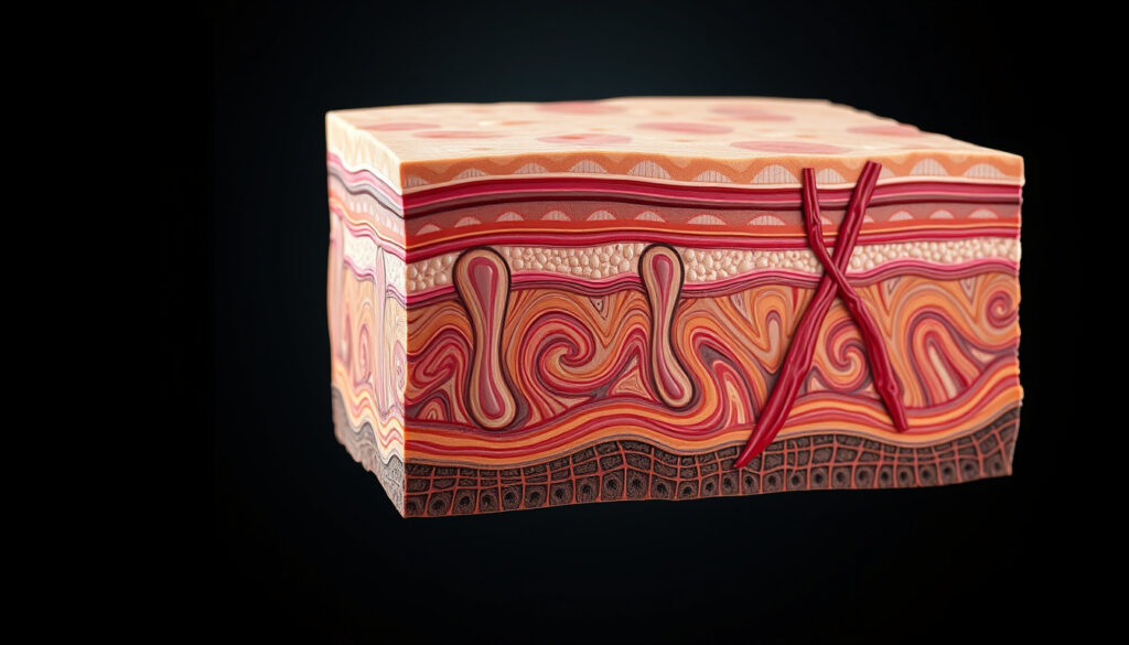
Layers of the Skin: Anatomy, Functions, and Common Questions
We don’t often give our skin enough credit for everything it does. It’s the largest organ in the body, acting as a barrier, a sensor, and an insulator all at once.
Main skin layers: 3 · Epidermis sublayers: 5 · Skin thickness range: 0.5–4 mm · Total skin surface area: about 1.5–2 m² · Skin weight: about 15% of body weight
Quick snapshot
- Skin has three primary layers: epidermis, dermis, hypodermis (NCBI Bookshelf)
- Epidermis contains no blood vessels (SEER Training Modules)
- Origin of the “7 layers of skin” claim in popular culture
- Which single layer is “most important” — all layers are interdependent
- Anatomy is stable from birth; no timeline in skin layer structure (StatPearls)
- Read on to understand the difference between 3, 4, 5, and 7 layer counts
- Learn how to remember the epidermal sublayers with a simple mnemonic
Five key facts, one pattern: each number refers to a different classification — main layers, epidermal sublayers, and marketing gloss.
| Label | Value |
|---|---|
| Number of main layers | 3 |
| Epidermal sublayers (thick skin) | 5 |
| Epidermal sublayers (thin skin) | 4 |
| Thickest layer | Dermis (most of skin’s thickness) |
| Layer with no blood vessels | Epidermis |
| Skin thickness range | 0.5–4 mm |
| Total skin surface area | about 1.5–2 m² |
| Skin weight | about 15% of body weight |
What Are the 3 Main Layers of the Skin?
Epidermis: the protective outermost layer
- Thin, avascular, stratified squamous epithelium (StatPearls)
- Produces melanin via melanocytes (SEER Training Modules)
- Acts as a barrier against pathogens and UV radiation (Osmosis)
The epidermis is the layer you see and touch. It contains no blood vessels — that’s why superficial cuts and scrapes don’t bleed.
Dermis: the structural support layer
- Contains collagen, elastin, blood vessels, and nerves (SEER Training Modules)
- Divided into papillary and reticular layers (NCBI Bookshelf)
- Supports temperature regulation and sensation (Osmosis)
This is where the real action happens — sweat glands, hair follicles, and nerve endings live here.
Hypodermis: the subcutaneous fat layer
- Lies deepest, attached to muscle and fascia (Cleveland Clinic)
- Contains adipose lobules, sensory neurons, and blood vessels (StatPearls)
- Provides insulation and energy storage (Osmosis)
Bottom line: The three-layer model (epidermis, dermis, hypodermis) is the standard anatomical answer. For medical accuracy, this is the system to use.
When a doctor tells you a cut is “only epidermal,” it means no bleeding and faster healing. When they say it reached the dermis, that’s when stitches may be needed. Knowing these layers directly affects how you treat wounds.
The implication: the three-layer model is the foundation for understanding skin injury and treatment.
What Are the 5 Layers of the Epidermis?
Stratum basale
- Deepest layer of the epidermis (StatPearls)
- Single row of cuboidal/columnar keratinocytes; site of cell division
- Contains melanocytes that produce melanin (SEER Training Modules)
Stratum spinosum
- Several cell layers thick; cells have spiny projections
- Provides strength and flexibility
Stratum granulosum
- 3-5 layers of flattened cells
- Cells produce keratohyalin granules that help form the barrier
Stratum lucidum
- Only present in thick skin (palms, soles)
- Clear, thin layer of dead keratinocytes
Stratum corneum
- Outermost layer; 15–30 layers of dead, flattened cells (Cleveland Clinic)
- Primary barrier against water loss and external insults
For students and clinicians, remembering the order from deep to superficial is a classic memorization hurdle — we’ll tackle that in a later section.
Thick skin (palms, soles) gets the extra stratum lucidum for added protection against friction. Thin skin, covering the rest of the body, is more flexible but less abrasion-resistant. Each design suits its location.
Knowing the sublayers helps clinicians assess wound depth and healing time.
Why Do Some Sources Say There Are 4 or 7 Layers?
4 layers in thin skin
- Thin skin lacks the stratum lucidum, leaving 4 epidermal sublayers
- About 90% of the body has thin skin
7 layers in cosmetic layering systems
- The “7 layers of skin” concept is not anatomical; it comes from cosmetic product layering
- Anatomical consensus remains 3 main layers (epidermis, dermis, hypodermis)
The implication: when you hear “7 layers,” you’re probably reading a beauty article, not a medical textbook. Stick with the histological count for accuracy.
Which Layer of Skin Does Not Bleed and Why?
Epidermis is avascular
- The epidermis contains no blood vessels (SEER Training Modules)
- All nutrients reach it via diffusion from the dermis
Bleeding indicates dermal injury
- Bleeding from a cut means the dermis has been reached (Cleveland Clinic)
- Burns that only damage the epidermis (first-degree) do not bleed
What this means: a scrape that oozes clear fluid but no blood is still epidermal. Once you see red, the dermis — and its capillaries — are involved.
How to Remember the 5 Layers of the Epidermis?
Mnemonic: “Come Lets Get Sun Burned”
- Corneum, Lucidum, Granulosum, Spinosum, Basale — from superficial to deep
- Alternative: “Before Signing, Get Legal Counsel” for deep to superficial
Visual memory techniques
- Draw a simple five-layer diagram and label each with a color
- Associate each layer with its key function: barrier (corneum), waterproof (lucidum), granular (granulosum), spine (spinosum), base (basale)
These memory aids are essential for students mastering skin anatomy.
What’s Confirmed and What’s Still Uncertain
Confirmed facts
- The skin has three main layers: epidermis, dermis, hypodermis (NCBI Bookshelf)
- Epidermis has 5 sublayers in thick skin and 4 in thin skin
- The dermis contains blood vessels, nerves, and appendages (SEER Training Modules)
- Hypodermis is primarily fat and connective tissue (Cleveland Clinic)
What’s unclear
- Where exactly the “7 layers of skin” myth originated — likely from cosmetic marketing
- Which single layer is “most important” — function depends on all layers working together
Skin is the largest organ, protecting from external elements.
– Cleveland Clinic
The skin has 3 layers—epidermis, dermis, and hypodermis—each with different structures and functions.
– NCBI Bookshelf (StatPearls)
All three layers contain specialized sensory nerve structures.
For anyone trying to make sense of skincare marketing or anatomy textbooks, the key is knowing which counting system is being used. Stick with the three-layer model for medical accuracy, and treat the 7-layer claim as a marketing artifact. For doctors and students, the five epidermal sublayers remain essential knowledge — and the mnemonic makes it manageable. The confusion around layer counts isn’t just cosmetic: it reflects real differences in how we classify the same organ depending on context.
my.clevelandclinic.org, youtube.com, histology.leeds.ac.uk, derickdermatology.com, medlineplus.gov, courses.lumenlearning.com, youtube.com, britishjournalofnursing.com
Frequently asked questions
How thick is human skin?
Human skin ranges from about 0.5 mm on the eyelids to 4 mm on the palms and soles. The average thickness across the body is about 1.5–2 mm.
What is the function of the stratum corneum?
The stratum corneum is the outermost layer of the epidermis, composed of dead keratinocytes. It serves as the primary barrier against water loss, pathogens, and physical abrasion.
Can skin layers repair after injury?
Yes. Epidermal cells regenerate from the stratum basale, while the dermis heals with scar tissue if the injury is deep. The hypodermis can also regenerate but slower.
What causes wrinkles in the skin?
Wrinkles result from loss of collagen and elastin in the dermis, combined with thinning of the epidermis and hypodermis. UV exposure and aging accelerate this.
Does the hypodermis contain nerves?
Yes, the hypodermis contains sensory neurons that detect pressure and temperature, along with blood vessels and hair follicle roots.
Why is the dermis called the “true skin”?
The dermis contains blood vessels, nerves, and glands — all the living components of skin. The epidermis is mostly dead cells, so anatomists often refer to the dermis as the “true skin”.
More related posts
 Holiday Inn Club Vacations: Resorts, Timeshares & Exit Info
 What Time Is Sunset Tonight in Ireland? Complete Guide
 Criminal Minds: Evolution Season 4 Premiere & Cast News
 Sean Combs Expedited Appeal: What It Means & Timeline
 What Time Is It in France? Current Time, Time Zone & DST
 NFL Strength of Schedule 2025: Easiest & Toughest Teams
 Fiber Internet Near Me – Find Providers, Check Availability
 A Little Life Summary – Spoiler-Free Plot, Characters, Themes